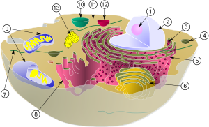
Wikipedia:Featured picture candidates/File:Biological cell.svg
Appearance

(2) nucleus
(3) ribosomes (little dots)
(4) vesicle
(5) rough endoplasmic reticulum (ER)
(6) Golgi apparatus
(7) Cytoskeleton
(8) smooth ER
(9) mitochondria
(10) vacuole
(11) cytoplasm
(12) lysosome
(13) centrioles within centrosome
- Reason
- Extremely high EV, possibly highest use I've ever seen, well done image, meets criteria. I can't believe no one found this gem for FP yet.
- Articles this image appears in
- over 100, including Cytoplasm, Lysosome, Organelle, Cell nucleus, pick any part of a cell and this is there, prominently.
- Creator
- MesserWoland and Szczepan1990
- Support as nominator -- Nezzadar ☎ 02:35, 20 October 2009 (UTC)
- Oppose, per criteria one, three, and lack of reliable sourcing. We have more technical illustrations of cells available. –blurpeace (talk) 23:53, 21 October 2009 (UTC)
- I strongly disagree with that statement. The guidelines are a minimum for FPs, and this image clearly meets those guidelines. I won't even address criteria one, its obvious that the image passes there. As for criteria three, the only possible reason to object is that the organelles are not labeled. This is addressed in the individual articles. Finally, I urge you to look at criteria number five. "A picture's encyclopedic value is given priority over its artistic value." I challenge you to find an image that has higher EV on more pages than this. Go ahead, spend six hours and find two images. Nezzadar ☎ 05:12, 20 October 2009 (UTC)
- Also, your arguement goes dangerously close to the fungi rationale. I.e. "we don't need another FP of a mushroom." This rationale has no weight here. If it meets FP requirements, it meets FP requirements, regardless of how many other similar images exist. Nezzadar ☎ 05:17, 20 October 2009 (UTC)
- The image lacks reliable sources. The ribosomes are entirely too simple, and since when are the contents of mitochondria shaped in the form of bundled shoelaces? Allow me to give you an illustration that is of featurable quality, File:Complete neuron cell diagram en.svg. The nomination pales in comparison.
Switching to strong oppose.–blurpeace (talk) 23:53, 21 October 2009 (UTC)- There we go, some arguments I can work with. First off, show me any image that has ribosomes as something other than dots. Umm really? Second, the mitochondria are not perfect, but the size, shape, and placement are. This is not nearly as technical as the neuron cell, it's designed to show size and placement in an easy to understand way. As for the sources, not much I can do to help you with that, except for the fact that the diagram looks like every diagram in every biology textbook short of med-school, so it can be seen as common knowledge to middle school graduates. Nezzadar ☎ 03:35, 22 October 2009 (UTC)
- The image lacks reliable sources. The ribosomes are entirely too simple, and since when are the contents of mitochondria shaped in the form of bundled shoelaces? Allow me to give you an illustration that is of featurable quality, File:Complete neuron cell diagram en.svg. The nomination pales in comparison.
- Also, your arguement goes dangerously close to the fungi rationale. I.e. "we don't need another FP of a mushroom." This rationale has no weight here. If it meets FP requirements, it meets FP requirements, regardless of how many other similar images exist. Nezzadar ☎ 05:17, 20 October 2009 (UTC)
- Comment: Could we please have a key in the image caption? Is currently pretty meaningless without it. J Milburn (talk) 11:24, 20 October 2009 (UTC)
- It is numbered in a number of articles. I copy pasta'd it. Noodle snacks (talk) 11:41, 20 October 2009 (UTC)
- Weak Support Seems widely used enough that mistakes have probably been ironed out. The use of numbers allows corrections etc. I'd like to see the diagram referenced for a full support. Noodle snacks (talk) 11:41, 20 October 2009 (UTC)
- How does high visibility absolve the illustration's errors? –blurpeace (talk) 23:56, 21 October 2009 (UTC)
- Simple, the points are numbered. Therefore any editor could correct mistakes. The high visibility makes the probability of such mistakes going unnoticed lower. Noodle snacks (talk) 04:13, 22 October 2009 (UTC)
- You'd have to wonder why most of our articles aren't up to GA yet. –blurpeace (talk) 18:20, 22 October 2009 (UTC)
- True, but this image does appear in a GA, and a FA for that matter. Noodle snacks (talk) 22:51, 22 October 2009 (UTC)
- Simple errors will be fixed over time in the way Noodle snacks describes- other problems (neutrality and such) will often not fix themselves, and it takes a lot more to expand and research than it does to correct and prettify. J Milburn (talk) 23:42, 22 October 2009 (UTC)
Update Tried labeling image in inkscape, failed, don't have time to learn the program. Nezzadar ☎ 16:36, 20 October 2009 (UTC)
- Comment Numbers don't cut it for me. Everything should be identified within the picture. Makeemlighter (talk) 03:38, 22 October 2009 (UTC)
- Weak oppose I can't imagine that this is the best way to label the organelles. I would have liked to see the names on the image itself. -- mcshadypl TC 04:21, 22 October 2009 (UTC)
- This is true. I tried editing it myself, but wound up losing the image when I redid the text. The redeeming value to this, however, is that the articles themselves will mention which number is relevant. In all likelyhood, this will be changed eventually though. Nezzadar ☎ 05:03, 22 October 2009 (UTC)
- Oppose, the picture is too simple. Comparing against Campbell's Biology (1995) shows it is lacking several important features of cells. First is the lack of 'free-roaming' ribosomes which strikes me as odd omission. Then is the lack of microtubules, microfilaments and other microstructures. The image also lacks peroxysomes, and the nucleus could have chromatine and nuclear pores depicted. The cell membrane is not identified. Lysosomes are not mere bubbles as this image implies, but rather have some inner structure. Also several animal cells have flagellums, which could be depicted. Headbomb {ταλκκοντριβς – WP Physics} 19:16, 22 October 2009 (UTC)
- Oppose Numbering is done for the purpose of allowing internationalisation, but is misguided imo since the use of SVGs makes it easy to translate such documents. The use of numbers rather than text labels leads to a lack of clarity and facilitates mistakes being made in the number-to-feature mapping. Papa Lima Whiskey (talk) 22:36, 22 October 2009 (UTC)
Not promoted --jjron (talk) 12:46, 27 October 2009 (UTC)
